
human anatomy laboratory manual with cat dissections
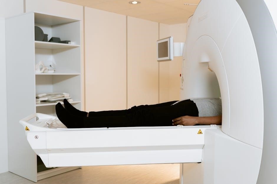
This laboratory manual seamlessly integrates the study of human anatomy with practical cat dissections‚ offering a comparative approach to understanding mammalian structures․
It provides detailed guidance for dissection activities‚ enhanced with full-color visuals‚ serving as a standalone resource or a companion to traditional anatomy coursework․
Students will rotate dissection roles‚ ensuring all members identify structures‚ while utilizing safety protocols and proper specimen handling techniques․
The manual emphasizes hands-on learning‚ fostering a deeper comprehension of anatomical relationships and physiological functions in both species․
It also acknowledges the emotional impact of loss‚ offering support for coping with grief‚ even in the context of animal studies․
Purpose of the Laboratory Manual
This manual’s core purpose is to deliver a comprehensive‚ hands-on exploration of mammalian anatomy‚ utilizing cat dissections as a key learning tool alongside human anatomy studies․
It aims to bridge theoretical knowledge with practical application‚ enabling students to visualize and understand complex anatomical structures․
The guide facilitates comparative analysis‚ highlighting similarities and differences between feline and human systems․
Furthermore‚ it cultivates essential laboratory skills‚ including proper dissection techniques‚ specimen handling‚ and accurate anatomical identification․
Ultimately‚ this resource prepares students for advanced studies in biology‚ medicine‚ and related fields․
Safety Precautions in the Anatomy Lab
Prioritizing safety is paramount in the anatomy lab․ Gloves and aprons must be worn at all times to prevent contact with specimens and potentially hazardous materials․
Work areas should be covered with multiple layers of newspaper to absorb fluids and facilitate cleanup․
Proper handling of dissection tools is crucial; exercise caution to avoid self-injury or damage to specimens․
Dispose of biological waste according to established laboratory protocols․
Maintain a clean and organized workspace‚ and report any accidents or spills to the instructor immediately․

Systematic Dissection Techniques
Effective dissection requires a methodical approach‚ carefully separating tissues to reveal underlying structures․ Rotate roles for hands-on experience‚ ensuring all students observe and identify anatomical features․
Tools and Equipment for Dissection
Essential tools for successful dissection include a sharp scalpel for precise incisions‚ forceps for grasping and manipulating tissues‚ and scissors for cutting through muscles and connective tissues․ Dissecting pins are crucial for securing specimens‚ while probes aid in identifying structures․
Safety glasses and gloves are mandatory for personal protection․ Aprons shield clothing from fluids․ Newspaper layers absorb spills‚ maintaining a clean workspace․ Proper tool handling and maintenance are vital for accurate and safe dissections․
Ensure all equipment is clean and in good working order before beginning the procedure․
Proper Handling of Specimens
Respectful handling of cat specimens is paramount․ Always maintain a professional demeanor and treat the animal with dignity․ Position the cat supine‚ securing limbs with pins․ Make shallow‚ controlled incisions to avoid damaging underlying structures․
Minimize tissue trauma during dissection․ Carefully reflect skin and muscle layers‚ identifying anatomical landmarks․ Dispose of biological waste properly‚ following laboratory protocols․
Remember‚ the specimen is a valuable learning tool; handle it with care and precision․

Skeletal System – Comparative Anatomy
This section explores feline and human skeletal similarities and differences‚ highlighting bone structure‚ joint types‚ and adaptations․ Comparative analysis reveals evolutionary relationships and functional variations․
Feline Skeletal Structure Overview
The cat skeleton exhibits a flexible spine enabling agility and a streamlined body for hunting․ Key features include a reduced clavicle‚ allowing greater shoulder movement‚ and digitigrade locomotion – walking on toes․
Observe the skull’s adaptations for carnivory‚ with prominent canines and a strong jaw․ The vertebral column comprises cervical‚ thoracic‚ lumbar‚ sacral‚ and caudal vertebrae․
Note the scapula’s position‚ facilitating powerful forelimb movements․ Limb bones demonstrate adaptations for speed and precision‚ crucial for predatory behavior․ Careful dissection reveals intricate skeletal details․
Human Skeletal System Overview
The human skeleton provides structural support‚ protects vital organs‚ and enables movement․ It consists of 206 bones‚ categorized into axial (skull‚ spine‚ ribs) and appendicular (limbs‚ girdles) divisions․
Observe the skull’s features adapted for bipedalism and large brain capacity․ The vertebral column supports upright posture and protects the spinal cord․
Note the clavicle’s role in shoulder stability and limb articulation․ Limb bones are adapted for weight-bearing and diverse movements․ Comparative analysis with feline skeletons highlights evolutionary adaptations․
Comparative Analysis: Cat vs․ Human Bones
Comparing feline and human skeletons reveals adaptations to different lifestyles․ Cats possess a more flexible spine for agility‚ while humans have a reinforced pelvis for bipedalism․
Observe the differences in limb proportions – cats have shorter limbs relative to body size‚ optimized for quadrupedal locomotion․
Note the presence of a clavicle in humans‚ providing greater shoulder range‚ whereas it’s reduced in cats․ Skull morphology differs‚ reflecting variations in sensory perception and feeding strategies․
Muscular System – Cat & Human
This section explores major muscle groups in both cats and humans‚ highlighting similarities and differences in muscle attachments and functions․
Comparative analysis reveals adaptations for locomotion‚ predation‚ and manipulation․
Major Muscle Groups in the Cat
Feline musculature exhibits distinct adaptations for agility and predatory behavior․ Key muscle groups include the powerful pectoralis and latissimus dorsi‚ crucial for forelimb movement and locomotion․
The biceps brachii and triceps brachii control elbow flexion and extension‚ while the gastrocnemius and soleus facilitate hindlimb propulsion․
Abdominal muscles provide core stability‚ and the diaphragm drives respiration․ Careful dissection reveals muscle origins‚ insertions‚ and functional relationships‚ demonstrating the cat’s remarkable physical capabilities․
Students will identify and analyze these muscles‚ comparing them to their human counterparts․
Major Muscle Groups in Humans
Human musculature is characterized by its complexity and capacity for diverse movements․ Prominent muscle groups include the pectoralis major‚ enabling arm adduction‚ and the latissimus dorsi‚ vital for back extension and arm rotation․
The biceps brachii and triceps brachii govern elbow flexion and extension‚ while the gastrocnemius and soleus are essential for plantar flexion of the foot․
Abdominal muscles provide core stability‚ and the diaphragm facilitates breathing․ Dissection and study reveal intricate muscle arrangements supporting human locomotion and function․
Muscle Tissue Types: Comparison
Human and feline muscle tissues share three primary types: skeletal‚ smooth‚ and cardiac․ Skeletal muscle‚ responsible for voluntary movement‚ exhibits striated patterns in both species‚ though fiber size may differ․
Smooth muscle‚ controlling involuntary functions like digestion‚ appears similar in both‚ lining internal organs․ Cardiac muscle‚ unique to the heart‚ demonstrates rhythmic contractions․
Comparative analysis reveals functional similarities‚ yet subtle structural variations reflecting species-specific adaptations․ Dissection aids in visualizing these distinctions and understanding muscle physiology․
Nervous System – Dissection & Identification
This section details cat brain dissection and human brain anatomy‚ alongside spinal cord and peripheral nerve identification‚ fostering comparative neurological understanding․
Cat Brain Dissection
Carefully dissecting the cat brain allows for direct observation of key structures‚ including the cerebrum‚ cerebellum‚ and brainstem․ Students will identify major lobes‚ fissures‚ and functional areas․
This hands-on experience complements the study of human brain anatomy‚ revealing fundamental similarities and differences in mammalian brain organization․
Precise identification of cranial nerves and associated pathways is crucial‚ enhancing understanding of sensory and motor functions․
Proper technique and respectful handling of the specimen are paramount throughout the dissection process;
Human Brain Anatomy – Overview
The human brain‚ a remarkably complex organ‚ is divided into distinct regions: the cerebrum‚ diencephalon‚ brainstem‚ and cerebellum․
Each area governs specific functions‚ from conscious thought and sensory perception to vital life support systems․
Detailed study of the cerebral cortex reveals specialized lobes responsible for motor control‚ sensory integration‚ and higher-order cognitive processes․
Comparative analysis with the cat brain highlights evolutionary adaptations and shared mammalian characteristics․
Understanding these structures is fundamental to comprehending neurological function and dysfunction․
Spinal Cord & Peripheral Nerves (Cat)
The feline spinal cord‚ protected within the vertebral column‚ serves as a crucial communication pathway between the brain and the body․
Careful dissection reveals its segmented structure‚ with dorsal and ventral roots giving rise to peripheral nerves․
These nerves innervate muscles and sensory receptors‚ enabling motor control and sensory perception throughout the cat’s body․
Observing the distribution of these nerves provides insight into the organization of the feline nervous system․
Comparative study with human anatomy reveals striking similarities in nerve pathways․

Digestive System – Comparative Study
Comparative analysis of cat and human digestive systems reveals shared functionalities—ingestion‚ digestion‚ absorption—but with anatomical variations reflecting dietary adaptations․
Dissection highlights structural differences․
Cat Digestive System Anatomy
The feline digestive tract begins with the mouth‚ leading to the esophagus and stomach—a muscular sac initiating protein digestion․ Further down‚ the small intestine‚ comprised of the duodenum‚ jejunum‚ and ileum‚ facilitates nutrient absorption․
The large intestine‚ including the cecum‚ colon‚ and rectum‚ reabsorbs water and forms feces․
Accessory organs—liver‚ gallbladder‚ and pancreas—contribute enzymes and bile for efficient breakdown․
Dissection reveals these structures‚ showcasing their arrangement and relative sizes within the abdominal cavity․
Human Digestive System Anatomy
The human digestive system mirrors the cat’s in basic function‚ starting with the mouth‚ pharynx‚ and esophagus delivering food to the stomach․ The small intestine—duodenum‚ jejunum‚ and ileum—is longer‚ maximizing nutrient absorption․
The large intestine‚ including the cecum‚ colon‚ rectum‚ and anus‚ processes waste․
Accessory organs like the liver‚ gallbladder‚ and pancreas aid digestion with enzymes and bile․
Comparative study highlights structural differences impacting digestive efficiency and dietary adaptations․
Comparative Analysis of Digestive Processes
Both cats and humans employ mechanical and chemical digestion‚ but differ in efficiency and enzyme specialization․
Cats‚ as carnivores‚ possess a shorter digestive tract optimized for protein breakdown‚ while humans‚ as omnivores‚ have a longer tract for plant matter․
Gastric acid strength and intestinal villi density vary‚ influencing nutrient absorption rates․
Comparing digestive secretions and gut motility reveals adaptations to respective diets․
This analysis underscores evolutionary pressures shaping digestive physiology in each species․
Respiratory System – Cat & Human
Comparative study reveals shared respiratory structures – lungs‚ trachea‚ diaphragm – yet differing lung capacities and branching patterns․
Dissection highlights feline lung lobulation‚ while human anatomy emphasizes alveolar density for efficient gas exchange․
Cat Respiratory System Dissection
Carefully expose the thoracic cavity‚ identifying the trachea‚ bronchi‚ and lungs․ Observe the distinct lobulation of feline lungs – typically three lobes on the right and two on the left – differing from human lungs․
Trace the path of airflow from the nasal cavity through the larynx and into the pulmonary system․ Dissect to reveal the diaphragm‚ crucial for breathing mechanics․
Note the cartilaginous rings supporting the trachea‚ preventing collapse during inhalation․ Examine the branching pattern of bronchioles within the lungs‚ facilitating gas exchange․
Proper technique ensures accurate observation and understanding of feline respiratory anatomy․
Human Respiratory System Anatomy
The human respiratory system comprises the nasal cavity‚ pharynx‚ larynx‚ trachea‚ bronchi‚ and lungs․ Unlike cats‚ human lungs exhibit three lobes on the right and two on the left․
Alveoli‚ tiny air sacs‚ facilitate gas exchange – oxygen intake and carbon dioxide removal – within the lungs․ The diaphragm‚ a muscular sheet‚ drives breathing by altering chest cavity volume․
Observe the branching pattern of the bronchial tree‚ ensuring efficient oxygen distribution․ Understanding these structures is vital for comprehending respiratory function and potential pathologies;
Detailed anatomical knowledge is crucial for comparative analysis․
Gas Exchange Mechanisms
Gas exchange‚ vital for life‚ occurs in the alveoli of both cats and humans․ Oxygen diffuses from the air into the blood‚ while carbon dioxide moves from the blood into the alveoli to be exhaled․
This process relies on partial pressure gradients and the thin walls of the alveoli and capillaries․ Hemoglobin within red blood cells efficiently transports oxygen throughout the body․
Compare the efficiency of gas exchange in both species‚ considering lung surface area and ventilation rates․ Understanding these mechanisms is fundamental to comprehending respiratory physiology․
Effective gas exchange sustains cellular respiration․

Circulatory System – Heart & Vessels
This section details cat heart dissection alongside human heart anatomy‚ identifying chambers‚ valves‚ and major blood vessels in both species for comparative study․
Students will learn about circulatory pathways․
Cat Heart Dissection
The cat heart dissection provides a hands-on opportunity to visualize cardiac anatomy․ Students will carefully dissect the pericardium to expose the heart‚ identifying the atria‚ ventricles‚ and major vessels like the aorta and vena cava․
Detailed observation of valve structures – tricuspid‚ mitral‚ pulmonary‚ and aortic – is crucial․
Precise identification of coronary arteries and veins demonstrates blood supply to the myocardium․
This practical experience builds a foundational understanding of mammalian heart structure‚ preparing students for comparative analysis with the human heart․
Human Heart Anatomy
The human heart‚ a four-chambered muscular organ‚ exhibits a complex structure vital for systemic circulation․ Detailed study focuses on the right and left atria‚ receiving deoxygenated and oxygenated blood respectively‚ and the powerful ventricles pumping blood to lungs and body․
Key features include the aortic and pulmonary valves‚ ensuring unidirectional blood flow․
Understanding the coronary circulation‚ supplying the heart muscle itself‚ is paramount․
Comparative analysis with the feline heart highlights both similarities and functional adaptations in mammalian cardiovascular systems․
Blood Vessel Identification (Cat & Human)
This section details identifying major arteries and veins in both cat and human specimens․ Key vessels include the aorta‚ vena cava‚ pulmonary artery/vein‚ and coronary vessels․
Dissection reveals the structural differences – vessel thickness‚ branching patterns – reflecting circulatory demands․
Students will trace blood flow pathways‚ correlating vessel structure with function․
Comparative analysis highlights similarities in basic vessel architecture‚ alongside species-specific adaptations․
Accurate identification is crucial for understanding cardiovascular physiology and potential pathologies․

Urogenital System – Comparative Anatomy
This manual explores feline and human urinary and reproductive systems‚ detailing anatomical structures and functional differences through dissection and comparative study․
Focus is placed on kidney structure‚ bladder‚ and associated reproductive organs․
Cat Urogenital System Dissection
Carefully dissect the feline abdominal cavity to expose the kidneys‚ ureters‚ urinary bladder‚ and associated reproductive structures․ Identify the renal cortex‚ medulla‚ and pelvis‚ tracing the ureters to the bladder․
Observe the bladder’s location and structure‚ noting the urethra’s path․ In males‚ locate the testes‚ epididymis‚ vas deferens‚ and prostate gland; in females‚ identify the ovaries‚ uterine horns‚ and vagina․
Document anatomical relationships and variations‚ comparing observations between individuals․ Proper handling and disposal of specimens are crucial throughout the dissection process․
Human Urogenital System Anatomy
The human urogenital system comprises the kidneys‚ ureters‚ bladder‚ and urethra – responsible for urine formation and excretion․ Reproductive organs include the testes (males)‚ ovaries (females)‚ and associated ducts and glands․
Kidneys filter blood‚ producing urine which travels via ureters to the bladder for storage․ The bladder expels urine through the urethra․ Male anatomy includes the prostate and seminal vesicles; female anatomy features the uterus and fallopian tubes․
Understanding these structures is vital for comprehending physiological processes and potential pathologies;
Functional Differences
Compared to cats‚ human kidneys exhibit a more developed cortical region‚ impacting filtration rates and concentrating abilities․ Human reproductive cycles are also distinct‚ featuring a menstrual cycle absent in most feline species․
Feline kidneys possess a prominent renal papilla‚ aiding in urine concentration‚ while human bladders have greater capacity․ Hormonal regulation of reproductive functions differs significantly‚ with humans exhibiting complex cyclical patterns․
These variations reflect adaptations to differing lifestyles and environmental pressures․
Endocrine System – Overview
This section details major endocrine glands in both cats and humans‚ exploring hormonal regulation․ Comparative aspects highlight similarities and differences in gland structure and function․
Understanding these systems is crucial for comprehending physiological processes․
Major Endocrine Glands (Cat & Human)
Both cats and humans possess key endocrine glands‚ including the pituitary‚ thyroid‚ adrenal‚ and pancreas․ The pituitary gland regulates other glands‚ while the thyroid controls metabolism․
Adrenal glands manage stress responses‚ and the pancreas regulates blood sugar․ Gonads (ovaries/testes) produce sex hormones․
Dissection reveals gland locations‚ while comparative study highlights structural variations․ Understanding these glands is vital for grasping hormonal control of bodily functions in both species․
Hormonal Regulation – Comparative Aspects
Hormonal regulation exhibits striking similarities between cats and humans‚ yet subtle differences exist․ Insulin‚ for example‚ regulates glucose in both‚ but response rates may vary․
Cortisol‚ the stress hormone‚ functions similarly‚ though feline stress responses can differ․ Comparative analysis reveals conserved pathways alongside species-specific adaptations․
This manual explores these nuances‚ emphasizing how hormonal imbalances impact physiology․ Understanding these comparative aspects enhances comprehension of endocrine function and disease processes․
Laboratory Exercises & Assessment
Assessment includes detailed dissection reports‚ graded on accuracy and completeness‚ alongside a practical exam testing anatomical identification skills․
Successful completion demonstrates mastery of comparative anatomy and dissection techniques․
Dissection Reports & Grading Criteria
Dissection reports require meticulous documentation of observations‚ including labeled diagrams and detailed descriptions of anatomical structures in both the cat and human․
Grading emphasizes accuracy in identification (40%)‚ clarity of presentation (30%)‚ and completeness of observations (30%)․
Reports must demonstrate a clear understanding of comparative anatomy‚ highlighting similarities and differences between feline and human systems․
Students are expected to rotate dissection roles‚ contributing individually to the report’s content and demonstrating collaborative learning․
Proper use of anatomical terminology is crucial for achieving a high score․
Practical Examination Format
The practical exam assesses your ability to identify anatomical structures on both preserved cat specimens and human anatomical models․
Stations will include dissections‚ requiring accurate labeling and functional explanations (60%)‚ and model identification (40%)․
You will demonstrate understanding of comparative anatomy‚ articulating key differences and similarities between feline and human systems․
Preparation involves thorough review of dissection guides‚ diagrams‚ and lecture material․
Rotating dissection roles during labs is excellent preparation for the exam’s hands-on component․