
ct scan ordering guide
CT Scan Ordering Guide: A Comprehensive Overview (Updated 12/19/2025)
Today‚ December 19th‚ 2025‚ presents a crucial need for informed CT scan ordering‚ balancing diagnostic benefits against potential long-term health consequences for patients.
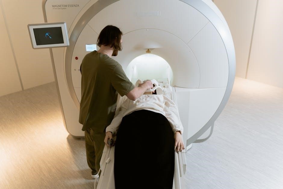
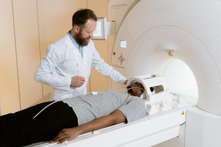
Computed Tomography (CT) scans are a vital diagnostic tool‚ employing X-rays to create detailed cross-sectional images of the body. These images allow clinicians to visualize bones‚ soft tissues‚ and blood vessels with remarkable clarity‚ aiding in the swift and accurate diagnosis of a wide spectrum of medical conditions.
However‚ the increasing utilization of CT scans‚ particularly in emergency room settings – rising from 7.8 million in 2007 to 16 million by 2022 – necessitates a careful consideration of their appropriate use. While invaluable for detecting injuries and illnesses‚ it’s essential to acknowledge emerging evidence suggesting potential long-term consequences associated with radiation exposure.

This guide aims to provide a comprehensive overview of the CT scan ordering process‚ emphasizing the importance of balancing the benefits of this powerful imaging modality with a commitment to patient safety and responsible radiation management. Understanding the risks and benefits is paramount for informed decision-making.
II. Understanding the Risks and Benefits
CT scans offer significant diagnostic advantages‚ enabling rapid and precise identification of internal injuries and diseases. This speed and accuracy can be life-saving in emergency situations‚ guiding critical treatment decisions. However‚ it’s crucial to acknowledge the inherent risks associated with this imaging technique.
The primary concern revolves around radiation exposure. While individual scan doses are generally low‚ cumulative exposure from multiple scans over a lifetime can potentially increase the risk of cancer. This is particularly relevant given the documented rise in CT scan usage‚ especially head CTs in emergency departments.
Furthermore‚ many CT scans utilize contrast dye to enhance image clarity. While generally safe‚ contrast dyes can cause allergic reactions‚ ranging from mild itching to severe anaphylaxis. Patients with pre-existing kidney conditions are also at increased risk of contrast-induced nephropathy. A thorough risk-benefit assessment is therefore essential before ordering a CT scan.
A. Radiation Exposure Concerns
Ionizing radiation‚ utilized in CT scans‚ carries a small but measurable risk of inducing cellular damage that could potentially lead to cancer development over time. The risk is cumulative‚ meaning it increases with each exposure throughout a patient’s life. While modern CT technology employs dose reduction techniques‚ the potential for harm remains a valid concern.
The increase in CT scan utilization is particularly noteworthy. Data reveals a substantial rise in head CT scans performed in emergency rooms – from 7.8 million in 2007 to 16 million between 2007 and 2022. This trend highlights the need for careful consideration of scan necessity.
Factors influencing radiation dose include scan parameters‚ patient size‚ and the area being imaged. Clinicians must prioritize the ALARA principle – “As Low As Reasonably Achievable” – when determining scan protocols‚ balancing image quality with minimizing radiation exposure. Patient education regarding these risks is also paramount.
B. Contrast Dye Risks & Allergies
CT scans often utilize contrast dyes‚ typically containing iodine‚ to enhance image clarity and improve diagnostic accuracy. However‚ these agents are not without potential risks. Allergic reactions‚ ranging from mild itching and hives to severe anaphylaxis‚ can occur‚ necessitating careful patient screening and preparedness for emergency intervention.
Patients with pre-existing kidney disease are particularly vulnerable to contrast-induced nephropathy (CIN)‚ a potential decline in kidney function following dye administration. Hydration protocols and alternative imaging modalities should be considered in these individuals.
A thorough patient history is crucial to identify allergies to iodine or shellfish – a common cross-reactivity. Clinicians must also inquire about any prior adverse reactions to contrast dye. Pre-medication with corticosteroids or antihistamines may be warranted in high-risk patients to mitigate potential allergic responses and ensure patient safety.
III. Clinical Indications for CT Scans
CT scans are invaluable diagnostic tools employed across a broad spectrum of clinical scenarios. Their rapid acquisition time and detailed anatomical visualization make them particularly useful in emergency medicine and critical care settings. However‚ appropriate utilization is paramount‚ guided by established clinical guidelines and a careful assessment of potential risks versus benefits.
Common indications include evaluating trauma‚ detecting internal bleeding‚ and assessing suspected fractures. CT scans are also crucial in diagnosing and monitoring various medical conditions affecting the brain‚ chest‚ abdomen‚ and pelvis. The increasing utilization of head CTs in emergency rooms‚ rising from 7.8 million in 2007 to 16 million by 2022‚ highlights their frequent application.
Careful consideration of alternative imaging modalities and adherence to the ALARA (As Low As Reasonably Achievable) principle are essential to minimize unnecessary radiation exposure while ensuring accurate and timely diagnoses.
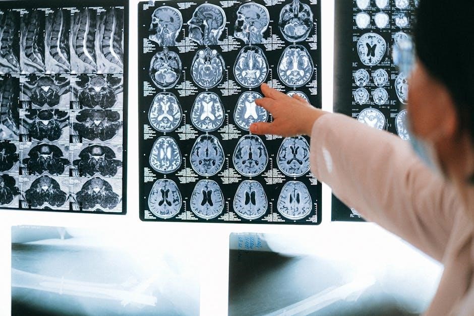
A. Neurological Applications
CT scans play a vital role in acute neurological assessments‚ particularly in emergency situations. They are exceptionally effective in rapidly identifying intracranial hemorrhages following head trauma‚ guiding immediate clinical interventions. The ability to quickly rule out or confirm bleeding is critical for patient management and prognosis.
Furthermore‚ CT scans are instrumental in stroke assessment‚ differentiating between ischemic and hemorrhagic strokes. This distinction is crucial as treatment strategies vary significantly depending on the stroke type. While advanced imaging like MRI offers greater detail‚ CT’s speed often makes it the initial choice.
The significant increase in head CT utilization – from 7.8 million in 2007 to 16 million in 2022 – underscores their frequent application in evaluating neurological symptoms. However‚ clinicians must weigh the benefits against radiation exposure‚ especially in younger patients.
Head Trauma & Bleeding
In cases of head trauma‚ a CT scan is often the first-line imaging modality due to its speed and accessibility. Its primary purpose is to rapidly identify intracranial hemorrhages – bleeding within the skull – which can be life-threatening; Detecting these bleeds quickly allows for prompt neurosurgical intervention‚ significantly improving patient outcomes.
CT scans excel at visualizing bony structures‚ making them ideal for identifying skull fractures that may accompany head injuries. They can also reveal signs of cerebral edema‚ or swelling of the brain‚ a common consequence of trauma. The information gleaned from a head CT directly influences treatment decisions‚ from conservative management to emergency surgery.

Given the rising number of head CTs performed in emergency rooms (increasing from 7.8 million in 2007 to 16 million by 2022)‚ careful consideration of the ALARA principle – As Low As Reasonably Achievable – regarding radiation dose is paramount.
Stroke Assessment
CT scans play a vital role in the rapid assessment of stroke‚ particularly in differentiating between ischemic and hemorrhagic stroke types. Identifying a hemorrhage is crucial‚ as treatment strategies differ dramatically. A CT scan can quickly rule out bleeding‚ allowing physicians to consider thrombolytic therapy – clot-busting drugs – for suspected ischemic strokes.
While CT scans may not always detect early ischemic changes‚ they serve as an essential initial step in the stroke workup; Newer CT perfusion techniques can provide more detailed information about blood flow to the brain‚ aiding in the identification of salvageable tissue. This information is critical for determining eligibility for advanced interventions like mechanical thrombectomy.
The speed of CT imaging is paramount in stroke care‚ as “time is brain.” Delays in diagnosis can lead to irreversible neurological damage. However‚ clinicians must remain mindful of the increasing utilization of head CTs and prioritize judicious use.
B. Thoracic Applications
CT scans are frequently utilized for evaluating a wide range of thoracic conditions. They offer detailed visualization of the lungs‚ mediastinum‚ and chest wall‚ proving invaluable in diagnosing acute and chronic ailments. Rapid assessment is particularly critical in emergency settings‚ such as suspected pulmonary embolism (PE).
For pneumonia and lung cancer screening‚ CT scans can detect subtle abnormalities often missed by chest X-rays. Low-dose CT scans are increasingly employed for lung cancer screening in high-risk individuals‚ such as long-term smokers. However‚ the benefits must be weighed against the potential risks of radiation exposure.
Furthermore‚ CT scans assist in evaluating chest trauma‚ identifying rib fractures‚ pneumothoraces‚ and internal organ injuries. Careful consideration of the ALARA principle is essential when ordering thoracic CTs‚ given the sensitivity of lung tissue to radiation.
Pulmonary Embolism (PE) Detection
CT Pulmonary Angiography (CTPA) is the gold standard for diagnosing pulmonary embolism (PE). This specialized CT scan utilizes intravenous contrast dye to visualize the pulmonary arteries‚ allowing for the detection of blood clots. Rapid and accurate PE diagnosis is crucial‚ as it significantly impacts patient outcomes.
CTPA offers high sensitivity and specificity in identifying both central and peripheral PEs. However‚ it’s essential to assess the patient’s risk factors and clinical probability of PE before ordering a CTPA‚ minimizing unnecessary radiation exposure. Alternative diagnostic pathways‚ like D-dimer testing‚ should be considered first in low-risk patients.
Careful evaluation of the CTPA images requires expertise in interpreting subtle findings‚ such as filling defects within the pulmonary arteries. Prompt reporting of results is vital to initiate appropriate anticoagulation therapy and prevent further complications.
Pneumonia & Lung Cancer Screening
CT scans play a vital role in diagnosing pneumonia‚ particularly when standard chest X-rays are inconclusive. CT imaging can identify the extent and severity of lung infiltrates‚ differentiating between various types of pneumonia and detecting complications like pleural effusions or abscesses.
Low-dose CT (LDCT) screening is recommended for high-risk individuals for lung cancer – typically those with a significant smoking history. LDCT can detect small nodules that may be indicative of early-stage lung cancer‚ potentially improving survival rates through timely intervention.
However‚ LDCT screening also carries risks‚ including false-positive results leading to unnecessary follow-up procedures and potential overdiagnosis. Careful patient selection and adherence to established screening guidelines are crucial to maximize benefits and minimize harms.
C. Abdominal & Pelvic Applications
CT scans are frequently utilized for evaluating acute abdominal pain‚ swiftly identifying conditions like appendicitis‚ diverticulitis‚ and bowel obstructions. Their ability to visualize the entire abdomen provides a comprehensive assessment‚ aiding in accurate diagnosis and guiding treatment decisions.
For suspected kidney stones‚ non-contrast CT scans are often the preferred imaging modality due to their high sensitivity and specificity. They can pinpoint the stone’s location‚ size‚ and potential obstruction of the urinary tract‚ informing management strategies.
CT scans also assist in the detection and staging of abdominal and pelvic cancers‚ evaluating tumor size‚ spread to lymph nodes‚ and involvement of adjacent organs. This information is critical for treatment planning and monitoring response to therapy.
Appendicitis Diagnosis
CT scans have become a cornerstone in the diagnostic pathway for suspected appendicitis‚ particularly in cases where the clinical presentation is atypical or inconclusive. They offer a rapid and reliable method for visualizing the appendix and surrounding tissues.
Key findings on CT indicative of appendicitis include appendiceal enlargement (diameter greater than 6mm)‚ periappendiceal inflammation‚ and the presence of an appendicolith – a calcified fecal mass within the appendix. These signs help differentiate appendicitis from other causes of abdominal pain.
However‚ judicious use is crucial‚ especially in younger patients‚ due to radiation exposure. Clinical assessment and alternative imaging modalities like ultrasound should be considered first when appropriate. CT remains invaluable when diagnosis is uncertain or complications are suspected.
Kidney Stone Evaluation
CT scans‚ specifically non-contrast helical CT‚ are the gold standard for evaluating suspected kidney stones (nephrolithiasis). Their high sensitivity and specificity allow for rapid and accurate detection of stones of varying sizes and compositions‚ even those radiolucent – meaning they don’t show up on standard X-rays.
The primary advantage of non-contrast CT is the avoidance of contrast dye‚ minimizing the risk of contrast-induced nephropathy‚ particularly in patients with pre-existing kidney disease. Stones appear as bright densities within the urinary tract‚ allowing for precise localization and size assessment.
CT also helps determine the presence of hydronephrosis – swelling of the kidney due to blockage – and identify any associated complications. While ultrasound can be used as a first-line option‚ CT provides a more comprehensive evaluation‚ especially for smaller stones or complex cases.
IV. The Ordering Process: A Step-by-Step Guide
Initiating a CT scan requires a systematic approach‚ prioritizing patient safety and justification. First‚ a thorough patient history and physical exam are essential to understand the clinical context and rule out contraindications. Document relevant allergies‚ especially to contrast dye‚ and assess renal function.
Next‚ clearly articulate the clinical question the CT scan aims to answer. This justification is crucial‚ adhering to the ALARA (As Low As Reasonably Achievable) principle regarding radiation exposure. Consider alternative imaging modalities before ordering a CT scan.

The order itself must include specific details: the type of CT scan (with or without contrast)‚ the area to be scanned‚ and any specific clinical concerns. Ensure proper communication with the radiology department and obtain informed consent from the patient‚ explaining the risks and benefits.
A. Patient History & Physical Exam
A comprehensive patient history is the cornerstone of appropriate CT scan ordering. Begin by meticulously documenting any prior imaging studies‚ focusing on recent scans and radiation exposure levels. Detailed allergy information is paramount‚ specifically inquiring about reactions to iodinated contrast media – even mild reactions require careful consideration.
Assess renal function through recent lab results (creatinine‚ eGFR) as compromised kidney function increases the risk of contrast-induced nephropathy. A thorough medical history should uncover pre-existing conditions like diabetes or cardiovascular disease‚ influencing risk stratification.
The physical exam should correlate with the clinical indication for the scan‚ guiding the radiologist’s focus. Document all pertinent findings‚ ensuring a clear link between the clinical presentation and the need for CT imaging. This detailed assessment minimizes unnecessary scans.
B. Justification for CT Scan – ALARA Principle
Adhering to the ALARA (As Low As Reasonably Achievable) principle is fundamental when considering a CT scan. Every request must be rigorously justified‚ demonstrating a clear clinical benefit that outweighs the inherent risks of radiation exposure. Avoid ordering CT scans solely for “rule-out” purposes; a specific clinical question must drive the decision.
Consider the increasing utilization rates – head CTs in emergency rooms rose significantly between 2007 and 2022 – and actively challenge the necessity of each scan. Explore alternative imaging modalities‚ like ultrasound or MRI‚ when clinically appropriate.
Document the clinical indication explicitly on the order form‚ justifying why CT is the preferred modality. This ensures transparency and accountability‚ promoting responsible imaging practices and minimizing patient exposure to ionizing radiation.
V. Alternative Imaging Modalities
Exploring alternatives to CT scans is crucial for minimizing radiation exposure‚ particularly given the documented increase in CT utilization. MRI offers a valuable alternative‚ especially when detailed soft tissue evaluation is needed and radiation is a concern. However‚ MRI access can be limited and is contraindicated in patients with certain metallic implants.
Ultrasound presents a first-line option for many clinical scenarios‚ offering real-time imaging without ionizing radiation. It’s particularly useful in evaluating abdominal and pelvic conditions‚ but image quality can be operator-dependent and limited by body habitus.
Careful consideration of each patient’s clinical presentation and available resources is essential when selecting the most appropriate imaging modality‚ always prioritizing patient safety and minimizing unnecessary radiation.
A. MRI vs. CT Scan – When to Choose Which
Choosing between MRI and CT scans requires careful clinical judgment. MRI excels in soft tissue detail‚ making it ideal for neurological assessments‚ ligament/tendon injuries‚ and spinal cord evaluations where radiation exposure is a concern. However‚ MRI is more time-consuming and expensive‚ and contraindicated for patients with certain metallic implants.
CT scans remain the gold standard for evaluating bone fractures‚ acute hemorrhage‚ and pulmonary emergencies due to their speed and accessibility. CT is also superior for imaging patients who cannot tolerate MRI or have contraindications.
Consider the clinical question: if soft tissue detail is paramount and radiation avoidance is key‚ MRI is preferred. For rapid assessment of bony structures or acute emergencies‚ CT remains the optimal choice.
B. Ultrasound as a First-Line Option
Ultrasound should be considered a valuable first-line imaging modality‚ particularly when evaluating certain clinical scenarios. It offers real-time imaging‚ lacks ionizing radiation‚ and is relatively inexpensive‚ making it suitable for initial assessments. For example‚ in suspected appendicitis‚ ultrasound can often visualize the appendix‚ reducing the need for immediate CT exposure.
Similarly‚ ultrasound is excellent for evaluating gallbladder disease‚ assessing fetal development during pregnancy‚ and guiding certain interventional procedures. However‚ ultrasound’s image quality is operator-dependent and can be limited by patient body habitus and bowel gas.
Prioritizing ultrasound when appropriate aligns with the ALARA principle‚ minimizing unnecessary radiation exposure while still providing valuable diagnostic information. It’s a crucial step in responsible CT scan ordering.
VI. Optimizing CT Scan Protocols
Optimizing CT protocols is paramount to delivering accurate diagnoses while minimizing patient radiation dose. Dose reduction techniques include utilizing automatic exposure control (AEC)‚ adjusting kVp and mAs settings based on patient size‚ and employing iterative reconstruction algorithms. These advancements significantly lower radiation without compromising image quality.
Contrast enhancement protocols also require careful consideration. Utilizing the lowest necessary contrast dose‚ tailoring the timing of contrast administration to the specific anatomy being imaged‚ and employing bolus tracking techniques can improve visualization while reducing potential adverse reactions.
Regular protocol review and standardization across institutions are essential. Implementing standardized protocols ensures consistent image quality and minimizes variability in radiation exposure‚ ultimately benefiting patient safety.
A. Dose Reduction Techniques

Implementing effective dose reduction techniques is crucial given the increasing utilization of CT scans and associated radiation exposure concerns. Automatic exposure control (AEC) dynamically adjusts radiation output based on patient size and attenuation‚ optimizing dose for each individual.
kVp modulation‚ lowering the peak voltage‚ can reduce patient dose without significantly impacting image quality‚ particularly in larger patients. Iterative reconstruction algorithms are sophisticated software tools that reduce image noise‚ allowing for lower radiation doses while maintaining diagnostic accuracy.
Shielding sensitive organs‚ like the thyroid and gonads‚ with appropriate lead shielding further minimizes unnecessary radiation exposure. Careful collimation to the area of interest and utilizing the shortest possible scan length also contribute to dose reduction.
B. Contrast Enhancement Protocols
Judicious use of intravenous contrast significantly enhances CT scan visualization of vascular structures and tissues‚ improving diagnostic confidence. Selecting the appropriate contrast agent – iodinated or gadolinium-based – depends on the clinical indication and patient factors‚ including renal function and allergy history.
Optimizing injection parameters‚ such as flow rate and contrast volume‚ is vital for achieving optimal enhancement. Timing the scan acquisition to coincide with peak arterial or venous enhancement maximizes visualization of specific structures. Dual-energy CT can characterize tissue composition and reduce contrast dose requirements.
Careful consideration of potential risks‚ including contrast-induced nephropathy and allergic reactions‚ is paramount. Hydration protocols before and after contrast administration can mitigate renal risks‚ while pre-medication may reduce allergic responses.
VII. Post-Scan Care & Follow-Up
Following a CT scan‚ diligent patient monitoring is essential‚ particularly for those receiving contrast. Observe for immediate adverse reactions like hives‚ itching‚ or difficulty breathing‚ requiring prompt intervention. Delayed reactions‚ though less common‚ can occur and necessitate reporting.

Hydration is crucial to help flush out the contrast dye‚ minimizing the risk of contrast-induced nephropathy‚ especially in patients with pre-existing renal issues. Clear communication of results to the patient is vital‚ explaining findings in understandable terms.
Appropriate follow-up depends on the scan’s findings; this may involve specialist referral‚ further imaging‚ or ongoing monitoring. Document all care and communication thoroughly‚ ensuring continuity of care and informed decision-making.
A. Monitoring for Adverse Reactions
Post-CT scan‚ immediate monitoring for adverse reactions is paramount‚ especially following contrast administration. Observe patients closely for at least 30 minutes for signs of allergic reactions – hives‚ itching‚ facial swelling‚ or respiratory distress. Prompt recognition and treatment with antihistamines or epinephrine may be necessary.
Delayed reactions‚ though rarer‚ can occur hours after the scan; educate patients to report any new symptoms. Assess renal function‚ particularly in at-risk individuals‚ as contrast can exacerbate pre-existing kidney issues. Document all observations meticulously‚ including vital signs and any interventions.
Ensure readily available emergency equipment and trained personnel are present during and immediately after the scan. Proactive monitoring minimizes potential complications and ensures patient safety.

B. Interpreting Results & Next Steps
Accurate interpretation of CT scan results requires a radiologist’s expertise‚ correlating imaging findings with the clinical context. Reports should be reviewed promptly‚ noting any incidental findings alongside the primary indication. Clear communication of results to the referring physician and patient is crucial.
Next steps depend entirely on the findings; normal scans may warrant clinical follow-up‚ while abnormal results necessitate further investigation or treatment. Consider the ALARA principle when planning follow-up imaging‚ exploring alternative modalities if appropriate. Document the rationale for any subsequent actions.
Patient education regarding results and follow-up plans is essential‚ addressing any concerns and ensuring understanding. Timely and appropriate action based on CT findings optimizes patient outcomes.